Summary
- Higher than 1 in 4 people die of sepsis throughout the world annually.
- Certain molecules of the pathogen bind to the receptors on specific immune cells, which become stimulated and trigger an acute inflammatory response, driven by pro-inflammatory and anti-inflammatory cytokines.
- Cytokines are the minor protein mediators formed by immune cells and comprise the interferons (IFN), interleukins (IL), chemokines, and tumor necrosis factors (TNF).
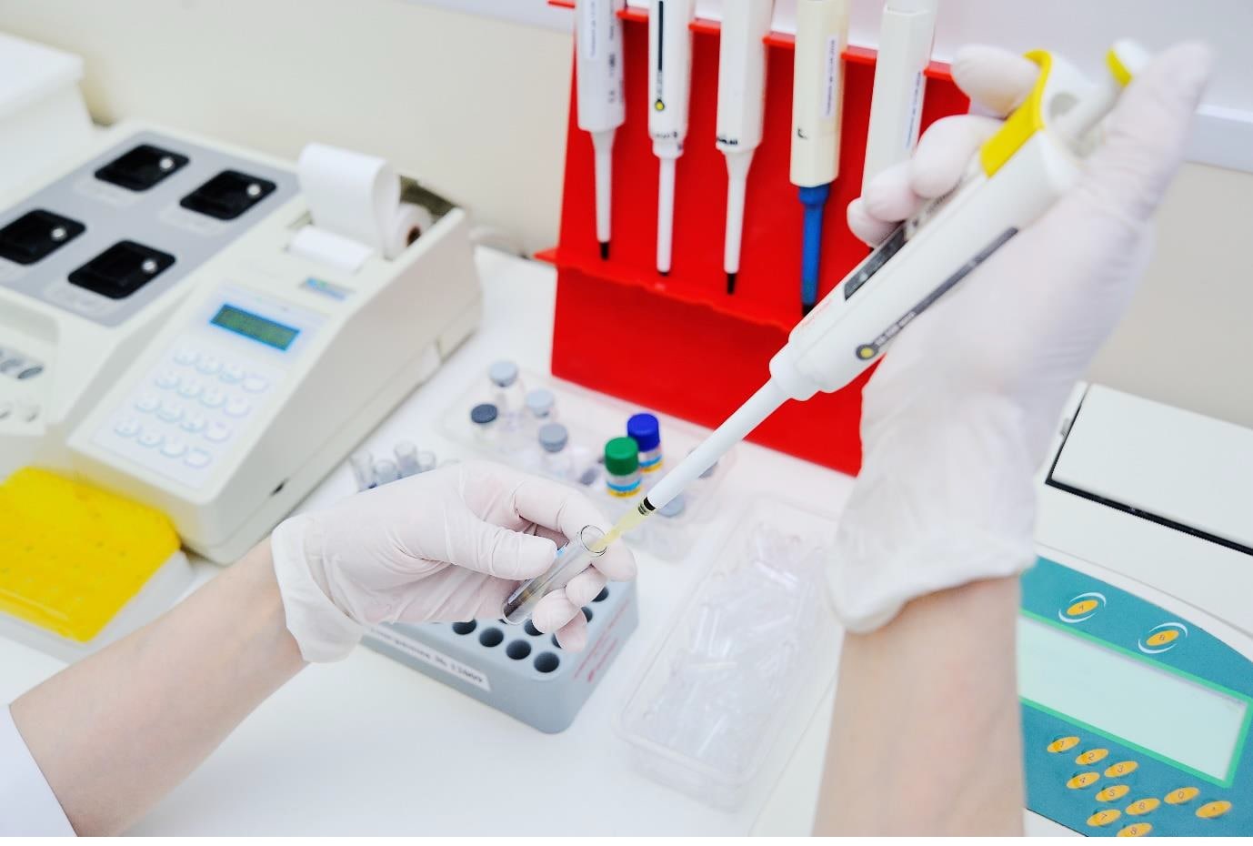
- Cytokine profiling techniques such as real-time polymerase chain reaction (RT PCR) have paved the way for the evaluation of several cytokines.
- Real-time PCR has evolved as an innovative technological advancement and is emerging with an essential role in the clinical diagnostics, prognostics, and research laboratories.
- The levels of certain cytokines found due to endothelial injury were significantly elevated in the initial acute phase in patients with sepsis than the control group, out of which IL-6 level had been elevated most significantly during the acute phase.
- A study’s findings showed that the IL-6 and PTX3 significantly declined in the patients who recovered and survived while the levels remained tall and even substantially elevated in the non-survivors, with IL-6 having a greater diagnostic and prognostic value.
- A decline in IL-6 levels is essentially linked with an improved prognosis and superior recovery, with its levels serving as the key predictor for clinical severity and fatal outcome.
- TNF-α is acknowledged as a vital pro-inflammatory cytokine that plays a role in producing cytokines such as IL-6, IL-8, and MCP-1, but its effect as a pro-inflammatory cytokine is partial or limited.
- The manifestation of biomarkers associated with the diagnosis or prognosis of sepsis can support clinicians and can improve patient care and the likelihood of survival.
Sepsis is one of the main reasons for death in the intensive care unit (ICU). More than 1 in 4 people die of sepsis across the globe annually. Certain molecules of the pathogen bind to the recognition receptors on specific immune cells for the pathogenesis of sepsis. The stimulated immune cells trigger an acute inflammatory response, which is driven by pro-inflammatory and anti-inflammatory cytokines, which play a role in the regulation of the inflammatory response.
In general, the term cytokine is used to describe the minor protein mediators formed by immune cells. Typically, cytokines comprise the interferons (IFN), interleukins (IL), chemokines, and tumor necrosis factors (TNF). It has been found that cytokines act on numerous cells and play a versatile role, including in inflammation and cellular differentiation. Cytokines are also considered to augment or inhibit the assembly of other cytokines.[1]
It has been established that levels of several cytokines are correlated with the prognosis and severity of acute inflammatory diseases, including sepsis.[2] However, the link between prognosis and severities of sepsis with cytokine networks has not yet been explained. Cytokine profiling techniques such as real-time polymerase chain reaction (RT PCR) have paved the way for evaluation of the several cytokines and expound on the status of cytokine networks.[3], [4]
What is Real-Time Polymerase Chain Reaction (RT PCR)?
Real-time PCR has evolved as an innovative technological advancement and is emerging with an essential role in the clinical diagnostics, prognostics, and research laboratories. Since it is capable of generating both quantitative and qualitative outcomes, Real-Time PCR is believed to be a quick and precise technique. This platform combines the response and demonstration of outcomes in a more rapid and accurate manner in comparison to the conventional PCR.[5]
In PCR, a DNA-polymerase enzyme that has a role in the replication of the cell’s genetic material is used to synthesize the specific DNA fragments. The advancement in the tools for DNA segment amplification has given rise to numerous benefits in the following fields.
- Gene analysis.
- Diagnosis of many genetic disorders.
- Detection of bacterial, viral, and fungal pathogens.[6]
Role of Interleukin 6 (IL-6) in the detection of sepsis
The levels of IL-6 (days 1, 2, 4, 6, 8, and 11), along with the other cytokines, have been shown to significantly increase compared to the controls, with its peak occurring on day 1, after which its levels gradually decline. In a 2018 study, it was stated that the levels of certain cytokines found as a result of endothelial injury were significantly elevated in the initial acute phase (day 1) in patients with sepsis than the control group. These include IL-1β, IL-6, IL-8, MCP-1, and PAI-1.[7]
Endothelial injury is greatly linked to the development of sepsis. This injury can proliferate the generation of cytokines, including IL-6, IL-8, and MCP-1. This points out that there may be a role of endothelial cell injury in the formation of the cytokine network consisting of IL-6, IL-8, and MCP-1 for the duration of the acute phase of sepsis. Additionally, the increase in IL-6, IL-8, and MCP-1 could initiate a positive feedback system by augmenting endothelial cells’ inflammatory response.[8]
Out of these cytokines, the level of IL-6 had been elevated most significantly during the acute phase. The striking concentration of IL-6 binds to the soluble form of the IL-6 receptor (IL-6R). This leads to the formation of the IL-6/IL-6R complex, which in turn pools with the cellular signal-transducing component – glycoprotein 130, including endothelial cells. As a result, IL-6 signal activation is elicited. Due to this process, endothelial injury is accelerated, leading to the accumulative generation of IL-6, IL-8, and MCP-1.[9]
In a prospective controlled study of 2019, it was reported that among the patients with septic shock at presentation, the preliminary levels of IL-6 within six hours of clinical diagnosis were 444.3 pg/mL in the recovered survivors and 7609.5 pg/mL in non-survivors (P = 0.05). The follow-up IL-6 levels in survivors and non-survivors within 24 hours after discharge were 21.5 and 9976.5 pg/mL, respectively (P < 0.001). These findings showed that the IL-6 and PTX3 significantly declined in the patients who recovered and survived (P < 0.001). The levels remained tall and even substantially elevated in the non-survivors (P = 0.03 and P = 0.009, respectively).
Some studies had shown that the diagnostic value of IL-6 was less than or equal to other specific cytokines. However, this study reported that serum levels of IL-6 and PTX3 were useful in identifying sepsis severity with optimum cut-off values. Furthermore, IL-6 is demonstrated to have greater diagnostic and prognostic value in comparison with PTX3, PCT, and CRP. IL-6 had been an independent risk factor for a 28-day death rate in people suffering from sepsis and septic shock.[10]
Essentially, a decline in IL-6 levels was linked with an improved prognosis and superior recovery. Hence, its levels were considered to be the key predictor for clinical severity and fatal outcome.
Role of Tumor Necrosis Factor Alpha (TNF-Α) in the detection of sepsis
TNF-α is acknowledged as a vital pro-inflammatory cytokine that plays a role in producing cytokines such as IL-6, IL-8, and MCP-1 along with IL-1β.9 However, the effects of TNF-α were not associated with the cytokines it gives rise to. This denotes the fact that TNF-α’s effect as a pro-inflammatory cytokine is partial or limited.
It was similarly seen that the pathogen infection response increased the pro-inflammatory cytokines including IL-6, IL-8, IL-18 as well as tumor necrosis factor-α (TNF-α) and anti-inflammatory cytokine (IL-10) in patients suffering from sepsis. However, the differences in the levels of TNF-α and certain other cytokines (IFN-α, IFN-γ, IL-12/IL-23p40, IL-17A, and IL-4) were not significant between the patients with sepsis and the controls.
Subsequently, high levels of serum IL-10 and TNF-alpha and an elevated IL-10 to TNF-alpha ratio were found to be connected with mortality, while increased levels of TNF-alpha, IL-6, among others, were identified in those patients having early hemodynamic deterioration.[10]
Another study conducted serial estimation of IL-6 and TNF-α in patients with sepsis. It was reported that the IL-6 levels declined from day 1 to 7 after admission in the survivor group. Also, the TNF-α level was substantially low on day 1 in the non-surviving female group.
Henceforth, the findings suggested that a descending trend in IL-6 levels was associated with better sepsis prognosis.[11]
Conclusion
The diagnostic procedure of sepsis has evolved with time and requires biomarkers that reflect infection, immune dysregulation, and organ dysfunction. The manifestation of biomarkers associated with the diagnosis or prognosis of sepsis can be supportive for clinicians and can improve patient care and the likelihood of survival.
Thus, the inflammatory cytokines circulating in serum have been recognized as mediators of impairment in sepsis patients.[12] Numerous studies have shown the upregulation of the inflammatory cytokines, including TNF-α and IL-6 in septic conditions as compared to the healthy individuals.
Subsequently, the systemic inflammatory response syndrome is considered to be linked with the progression of sepsis.[13] Hence, identification of inflammatory markers is taken as a means to estimate the prognosis of sepsis.
References:
1. Amarasekara DS, Yu J, Rho J. Bone Loss Triggered by the Cytokine Network in Inflammatory Autoimmune Diseases. J Immunol Res. 2015;2015:832127. doi:10.1155/2015/832127
2. Pierrakos C, Vincent JL. Sepsis biomarkers: a review. Crit Care. 2010;14(1):R15. doi:10.1186/cc8872
3. Mera S, Tatulescu D, Cismaru C, et al. Multiplex cytokine profipng in patients with sepsis. APMIS. 2011;119(2):155-163. doi:10.1111/j.1600-0463.2010.02705.x
4. Bozza FA, Salluh JI, Japiassu AM, et al. Cytokine profiles as markers of disease severity in sepsis: a multiplex analysis. Crit Care. 2007;11(2):R49. doi:10.1186/cc5783
5. Morillo JM, Lau L, Sanz M, Herrera D, Silva A. Quantitative real-time PCR based on single copy gene sequence for detection of Actinobacillus actinomycetemcomitans and Porphyromonas gingivaps. J Periodontal Res. 2003;38(5):518-524. doi:10.1034/j.1600-0765.2003.00684.x
6. Atkins SD, Clark IM. Fungal molecular diagnostics: a mini review. J Appl Genet. 2004;45(1):3-15.
7. Matsumoto, H., Ogura, H., Shimizu, K. et al. The cpnical importance of a cytokine network in the acute phase of sepsis. Sci Rep 8. 2018:13995. doi:10.1038/s41598-018-32275-8
8. Werle M, Schmal U, Hanna K, Kreuzer J. MCP-1 induces activation of MAP-kinases ERK, JNK and p38 MAPK in human endothepal cells. Cardiovasc Res. 2002;56(2):284-292. doi:10.1016/s0008-6363(02)00600-4
9. Tanaka T, Narazaki M, Kishimoto T. Immunotherapeutic imppcations of IL-6 blockade for cytokine storm. Immunotherapy. 2016;8(8):959-970. doi:10.2217/imt-2016-0020
10. Song J, Park D.W, Moon S, Cho H-J, Park J H, Seok H, Choi W S. Diagnostic and prognostic value of interleukin-6, pentraxin 3, and procalcitonin levels among sepsis and septic shock patients: a prospective controlled study according to the Sepsis-3 definitions. BMC Infect Dis. 2019;19:968. doi:10.1186/s12879-019-4618-7
11. Chaudhry H et al. Role of cytokines as a double-edged sword in sepsis. In Vivo. 2013;27(6):669-684.
12. de Andrade JAA, Gayer CRM, Nogueira NPA, et al. The effect of thiamine deficiency on inflammation, oxidative stress and cellular migration in an experimental model of sepsis. J Inflamm (Lond). 2014;11:11. Pubpshed 2014 Apr 24. doi:10.1186/1476-9255-11-11
13. Chou HL, Han ST, Yeh CF, et al. Systemic inflammatory response syndrome is more associated with bacteremia in elderly patients with suspected sepsis in emergency departments. Medicine (Baltimore). 2016;95(49):e5634. doi:10.1097/MD.0000000000005634